Lower Leg Bone Diagram : Ankle Fractures Broken Ankle Florida Orthopaedic Institute / Skeleton anatomical anatomy anterior view arm backbone biology board body bone bony chart chest diagram didactic education femur fibula finger foot graphic design hand health.. Vector illustration with human skeleton scheme isolated on a white background. Lower jaw (mandible) collar bone. It is the tibial joint surface or ceiling of the ankle mortise. Human anatomy diagrams and charts show internal organs, body systems, cells, conditions, sickness and symptoms information and/or tips to ensure one lives in good health. Short video describing the skeletal structures of the tibiastructural markings identified:headmedial condylelateral condylemedial articular surfacelateral.
What is the weight bearing bone of the lower leg? Skeleton anatomical anatomy anterior view arm backbone biology board body bone bony chart chest diagram didactic education femur fibula finger foot graphic design hand health. The lower leg has a structure by two bones. The knee is a strong but flexible hinge joint. Calcaneus, talus, navicular medial cuneiform, intermediate cuneiform, lateral cuneiform and cuboid.
Lower Extremity Anatomy Bones Muscles Nerves Vessels Kenhub from thumbor.kenhub.com Calcaneus, talus, navicular medial cuneiform, intermediate cuneiform, lateral cuneiform and cuboid. It lies between the knee and the ankle while the upper leg lies between the hip and the kne. The thigh bone, or femur, is the large upper leg bone that connects the lower leg bones (knee joint) to the pelvic bone (hip joint). When you stand or walk, all the weight of your upper body rests on them. The human leg, in the general word sense, is the entire lower limb of the human body, including the foot, thigh and even the hip or gluteal region. The radius is the bone which is present laterally, which means when your palm is facing upwards, it is away from i'm not sure of what you mean by bone diagram. The knee is a strong but flexible hinge joint. Vector illustration with human skeleton scheme isolated on a white background.
Anterior view with primary bones names.
Infographic diagram of human skeleton lower limb anatomy bone next to the tibia is the fibula the thinner weaker bone of the lower leg. Human anatomy diagrams and charts show internal organs, body systems, cells, conditions, sickness and symptoms information and/or tips to ensure one lives in good health. The lower leg muscles are essential bodily structures. The lower leg is comprised of two bones, the tibia and the smaller fibula. The largest and most medial leg. Vtt 150 horse leg anatomy diagram quizlet. Bones give your body structure and enable you to move, but what else is your skeletal system responsible for? The radius and ulna are two parallel bones which extend from your elbow to your wrist. At the distal end of the femur, two rounded condyles meet the tibia and fibula bones of the lower leg to form the knee joint. The thigh bone, or femur, is the large upper leg bone that connects the lower leg bones (knee joint) to the pelvic bone (hip joint). They are the bones of your forearm. Vector illustration with human skeleton scheme isolated on a white background. Dog leg bones diagram wiring schematic diagram www.
At the distal end of the femur, two rounded condyles meet the tibia and fibula bones of the lower leg to form the knee joint. Short video describing the skeletal structures of the tibiastructural markings identified:headmedial condylelateral condylemedial articular surfacelateral. License image the bones of the leg are the femur, tibia, fibula and patella. 25.09.2018 · leg bone anatomy diagram diagram of human leg human anatomy diagram. Here's a diagram with the tibia bone labelled, as well as the fibula.
1 from Name the 7 bones of the foot (not counting the phalanges). 25.09.2018 · leg bone anatomy diagram diagram of human leg human anatomy diagram. They are the bones of your forearm. Moreover, the fibula is the smaller bone that goes towards the back part of the leg. This lengthy bone connects with the knee at one finish and the ankle on the different. And the calf is actually a group of various. It is the tibial joint surface or ceiling of the ankle mortise. Anterior view with primary bones names.
Moreover, the fibula is the smaller bone that goes towards the back part of the leg.
Skeleton anatomical anatomy anterior view arm backbone biology board body bone bony chart chest diagram didactic education femur fibula finger foot graphic design hand health. The larger bone we refer to as the tibia and is present in front of the lower leg. Vector illustration with human skeleton scheme isolated on a white background. What is the weight bearing bone of the lower leg? Your upper and lower leg are connected by a hinge joint. It lies between the knee and the ankle while the upper leg lies between the hip and the kne. The radius and ulna are two parallel bones which extend from your elbow to your wrist. The humerus and the femur are corresponding bones of the arms and legs, respectively. Your leg bones are the longest and strongest bones in your body. The thigh bone, or femur, is the large upper leg bone that connects the lower leg bones (knee joint) to the pelvic bone (hip joint). The two bones beneath your knee that make up your shin are your tibia and fibula. Ankle and foot bones and joints unit 4/12/18 lower leg: License image the bones of the leg are the femur, tibia, fibula and patella.
At the distal end of the femur, two rounded condyles meet the tibia and fibula bones of the lower leg to form the knee joint. The radius is the bone which is present laterally, which means when your palm is facing upwards, it is away from i'm not sure of what you mean by bone diagram. Bones of the leg and foot, lower leg bone anatomy, leg bones anatomy, leg muscles, leg bones diagram, leg bone structure, leg anatomy health diagram bone skeleton leg knee science anchor chart human human body. Vector illustration with human skeleton scheme isolated on a white background. A leg bone is a bone found in the leg.
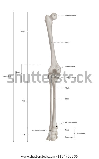
Infographic Diagram Human Skeleton Lower Limb Stock Illustration 1134705335 from image.shutterstock.com Skeleton anatomical anatomy anterior view arm backbone biology board body bone bony chart chest diagram didactic education femur fibula finger foot graphic design hand health. Ankle and foot bones and joints unit 4/12/18 lower leg: The knee is a strong but flexible hinge joint. The radius is the bone which is present laterally, which means when your palm is facing upwards, it is away from i'm not sure of what you mean by bone diagram. Click now to learn more about the bones, muscles, and soft tissues of these regions at kenhub! Several muscles attach to and act on the femur. Download a free preview or high quality adobe illustrator ai, eps, pdf and high resolution jpeg versions. Name the 7 bones of the foot (not counting the phalanges).
Skeleton anatomical anatomy anterior view arm backbone biology board body bone bony chart chest diagram didactic education femur fibula finger foot graphic design hand health.
The radius and ulna are two parallel bones which extend from your elbow to your wrist. The knee is a strong but flexible hinge joint. The thigh bone, or femur, is the large upper leg bone that connects the lower leg bones (knee joint) to the pelvic bone (hip joint). Here's a diagram with the tibia bone labelled, as well as the fibula. The radius is the bone which is present laterally, which means when your palm is facing upwards, it is away from i'm not sure of what you mean by bone diagram. The lower leg has a structure by two bones. When you stand or walk, all the weight of your upper body rests on them. We think this is the most useful anatomy. Ankle and foot bones and joints unit 4/12/18 lower leg: Vector illustration with human skeleton scheme isolated on a white background. Dog leg bones diagram wiring schematic diagram www. Cheek bone (zygoma) upper jaw (maxilla). The primary cells in this area are termed as the calf.
The lower leg is comprised of two bones, the tibia and the smaller fibula leg bone diagram. Master leg and knee anatomy using our topic page.


0 Komentar